
Introduction to Peroxisomal Disorders
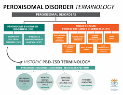
Peroxisomes are essential intracellular organelles involved in numerous metabolic pathways, particularly lipid metabolism and reactive oxygen species detoxification. Modern understanding of peroxisomal diseases classifies them into two major categories: peroxisome biogenesis disorders (PBDs) and single (isolated) enzyme deficiencies.
This classification has evolved over several decades of biomedical research. Although peroxisomes were first identified more than half a century ago, their association with human disease was not established until 1973, when Sidney Goldfischer observed the absence of peroxisomes in liver and kidney tissues from patients with Zellweger syndrome.
Subsequent milestones included:
- The discovery of the first diagnostic biomarker in 1984
- Identification of the first causative gene mutation in 1992
Interestingly, the clinical syndromes associated with PBDs were described well before their molecular and biochemical mechanisms were understood. This makes PBDs historically significant, as they represent one of the earliest examples of developmental malformations caused by metabolic dysfunction.
Classification of Peroxisome Biogenesis Disorders
PBDs encompass a spectrum of genetic conditions characterized by defective peroxisome assembly and function. The four classical syndromes include:
- Zellweger syndrome (ZS)
- Neonatal adrenoleukodystrophy (NALD)
- Infantile Refsum disease (IRD)
- Rhizomelic chondrodysplasia punctata (RCDP)
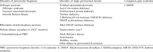
Advances in biochemical and molecular biology revealed that many patients with similar clinical features actually have distinct underlying mechanisms. Some present with defects in a single enzyme rather than global peroxisome dysfunction.
Today, at least 13 complementation groups have been identified, each associated with mutations in specific PEX genes, which encode proteins essential for peroxisome assembly (peroxins).
Clinical Phenotypes of PBD
Zellweger Syndrome Spectrum (ZSS)
The Zellweger spectrum disorders represent a continuum ranging from severe to milder phenotypes.
1. Classic Zellweger Syndrome (ZS)
ZS is the most severe form and presents at birth with multisystem involvement:
Clinical features:
- Distinct craniofacial abnormalities (high forehead, flattened midface)
- Severe hypotonia (muscle weakness)
- Seizures and feeding difficulties
- Absence of reflexes
- Profound neurological impairment
ZS affects nearly all organ systems due to the fundamental role of peroxisomes:
- Neurological system: neuronal migration defects, polymicrogyria
- Ocular system: cataracts, glaucoma, retinal abnormalities
- Hepatic system: hepatomegaly and liver dysfunction
- Renal system: cyst formation
- Skeletal system: chondrodysplasia punctata
The disease is typically fatal within the first year of life, with no significant developmental progress.
2. Neonatal Adrenoleukodystrophy (NALD) and Infantile Refsum Disease (IRD)
These conditions represent milder variants within the ZSS continuum.
Characteristics:
- Partial psychomotor development (e.g., sitting, walking)
- Variable neurological decline
- Less pronounced craniofacial abnormalities
- Possible development of leukodystrophy (myelin degeneration)
Unlike ZS:
- Some patients survive into adolescence or adulthood
- Clinical presentation is more heterogeneous
Common complications include:
- Sensorineural hearing loss
- Retinitis pigmentosa
- Progressive neurological deterioration
3. Rhizomelic Chondrodysplasia Punctata (RCDP)
RCDP is a distinct PBD with a unique phenotype.
Major features:
- Shortening of proximal limbs (rhizomelia)
- Skeletal abnormalities and calcifications
- Severe growth and developmental delay
- Congenital cataracts
- Neurological impairment
Additional complications:
- Respiratory issues
- Seizures
- Reduced life expectancy (variable)
Unlike ZSS, RCDP primarily involves defects in plasmalogen synthesis, which are critical phospholipids in cell membranes, especially in the nervous system.
Biochemical Basis and Diagnosis
Diagnostic Biomarkers
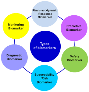
PBDs are diagnosed using biochemical markers in blood and tissues:
ZSS Profile:
- Elevated very long-chain fatty acids (VLCFA)
- Increased bile acid intermediates
- Reduced plasmalogens
RCDP Profile:
- Normal VLCFA levels
- Markedly reduced plasmalogens
Additional markers include:
- Phytanic acid
- Pristanic acid
- Pipecolic acid
However, biochemical overlap exists, making cellular and molecular testing essential.
Cellular and Molecular Diagnostics
Cultured fibroblasts are widely used to:
- Measure peroxisomal β-oxidation activity
- Assess plasmalogen synthesis
- Evaluate enzyme function
Immunocytochemistry allows visualization of peroxisomes:
- Reduced or absent peroxisomes
- Presence of “peroxisomal ghosts” (membrane remnants)
Genetic testing identifies mutations in PEX genes, confirming diagnosis and enabling:
- Carrier detection
- Prenatal diagnosis
Molecular Pathogenesis: Role of PEX Genes
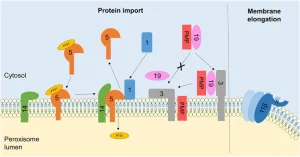
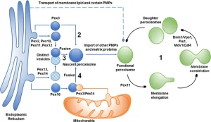
To date, mutations in at least 13 PEX genes have been linked to PBDs. These genes encode proteins essential for:
- Peroxisome membrane formation
- Protein import into peroxisomes
- Recycling of receptor proteins
Genes:
- PEX1, PEX6, PEX26 → receptor recycling
- PEX5, PEX7 → protein import receptors
- PEX2, PEX10, PEX12 → membrane translocation
- PEX3, PEX16, PEX19 → membrane assembly
Among these, PEX1 mutations account for ~70% of cases, making it the most common genetic cause.
Metabolic Pathways Affected in PBD
1. β-Oxidation of Fatty Acids
Peroxisomes degrade very long-chain fatty acids. Defects lead to toxic accumulation, particularly in:
- Brain
- Liver
- Adrenal glands
This contributes to neurological and systemic damage.
2. α-Oxidation of Phytanic Acid
This pathway prevents accumulation of phytanic acid from dietary sources.
Defects can lead to symptoms such as:
- Retinal degeneration
- Neuropathy
- Ataxia
3. Plasmalogen Biosynthesis
Plasmalogens are essential membrane lipids, especially in:
- Myelin
- Cardiac tissue
Deficiency contributes to:
- Neurological dysfunction
- Skeletal abnormalities (notably in RCDP)
4. Pipecolic Acid Metabolism
Although useful diagnostically, its exact role in disease progression remains unclear.
Advanced Concepts: Mosaicism and Temperature Sensitivity
Peroxisomal Mosaicism
Refers to variability in peroxisome function:
- Between tissues
- Between cells within the same tissue
This complicates diagnosis and phenotype prediction.
Temperature Sensitivity
Some mutations exhibit improved function at lower temperatures (e.g., 30°C), suggesting:
- Protein misfolding mechanisms
- Potential for pharmacological chaperone therapies
Prenatal Diagnosis and Genetic Counseling
Due to the severity of PBDs, prenatal testing is crucial.
Available methods:
- Biochemical assays (CVS or amniotic fluid)
- DNA-based mutation analysis
- Preimplantation genetic diagnosis (PGD)
Imaging techniques such as fetal MRI and ultrasound can detect:
- Brain abnormalities
- Skeletal defects
Current and Emerging Therapeutic Strategies
At present, treatment remains supportive, focusing on symptom management:
- Seizure control
- Nutritional support
- Vision and hearing interventions
Experimental approaches include:
- Dietary restriction (e.g., phytanic acid)
- DHA supplementation
- Bile acid therapy
- Plasmalogen precursors
- Liver transplantation (limited cases)
Future directions:
- Small molecule therapies to improve protein folding
- Gene-based therapies
- Targeted metabolic correction
Conclusion
Peroxisome biogenesis disorders represent a complex group of inherited metabolic diseases with profound developmental and systemic consequences. Advances in molecular genetics and biochemical diagnostics have significantly improved our understanding of these disorders, revealing intricate links between metabolism and human development.
Despite the absence of curative therapies, ongoing research into peroxisomal biology, gene function, and metabolic pathways offers promising avenues for future treatment strategies. Continued integration of clinical, biochemical, and genetic data will be essential for improving diagnosis, management, and patient outcomes.